|
 
 |
| ORIGINAL ARTICLE |
|
| Year : 2013 | Volume
: 1
| Issue : 1 | Page : 11-15 |
|
To establish the validity of dental age assessment using Nolla's method on comparing with skeletal age assessed by hand-wrist radiographs
Sachan Kiran1, Vijay Prakash Sharma2, Pradeep Tandon2, Tripti Tikku1, Snehlata Verma3, Kamna Srivastava1
1 Departments of Orthodontics and Dentofacial Orthopedics, BBDCODS, Lucknow, Uttar Pradesh, India
2 Department of Orthodontics, KGMC, Lucknow, Uttar Pradesh, India
3 Department of Orthodontics and Dentofacial Orthopedics, Uttar Pradesh Dental College and Research Centre, Lucknow, Uttar Pradesh, India
| Date of Web Publication | 18-May-2013 |
Correspondence Address:
Sachan Kiran
2/894 Sec-H Kursi Road, Jankipuram, Lucknow, Uttar Pradesh
India
 Source of Support: None, Conflict of Interest: None  | Check |

Background: Skeletal age assessment by hand-wrist radiographs has been found to correlate significantly with the growth status of an individual, but has a known drawback in the form of extra-radiograph and high dose of radiation exposure in comparison to periapical X-rays used commonly in dentistry. Aims and Objectives: The purpose of the study was to assess skeletal age using hand-wrist radiographs and to find the correlation amongst the skeletal, dental, and chronological ages. Materials and Methods : Ninety Indian healthy children in the age group 9-13 years, comprising equal number of males and females, were included in the study. The children were radiographed for hand-wrist of the right hand and intraoral periapical X-ray for right permanent maxillary and mandibular canine. Results: There was high correlation between skeletal maturation indicator and canine calcification stages for both male and female children (0.635, 0.891). Conclusion: Females were more advanced in skeletal maturation than males. Chronological age showed inconsistent correlation with dental and skeletal ages. It was concluded that canine calcification stages can also be used for assessing skeletal maturity. Keywords: Chronological age, canine, dental age, hand-wrist, skeletal age
How to cite this article:
Kiran S, Sharma VP, Tandon P, Tikku T, Verma S, Srivastava K. To establish the validity of dental age assessment using Nolla's method on comparing with skeletal age assessed by hand-wrist radiographs. J Orthod Res 2013;1:11-5 |
How to cite this URL:
Kiran S, Sharma VP, Tandon P, Tikku T, Verma S, Srivastava K. To establish the validity of dental age assessment using Nolla's method on comparing with skeletal age assessed by hand-wrist radiographs. J Orthod Res [serial online] 2013 [cited 2017 Apr 12];1:11-5. Available from: http://www.jorthodr.org/text.asp?2013/1/1/11/112250 |
| Introduction | |  |
Growing individuals not only differ in the timing of the maturational events, but also in the sequence of these events. The developmental status of a child can be accessed from various parameters such as height, weight, chronological age, secondary sexual characteristics, skeletal age, and dental age. [1] Skeletal age has been considered the most reliable method to assess the developmental status. [2],[3] The hand-wrist radiograph is commonly used for skeletal development assessment. The most frequently used method to evaluate skeletal age from hand-wrist radiographs is the Atlas More Details of Greulich and Pyle. [4] Dental age estimation is based upon the rate of development and calcification of tooth buds and the progressive sequence of their eruption in the oral cavity. Several methods have been developed to assess the dental age according to the degree of calcification observed in permanent teeth. One such widely used method is that given by Nolla. [5] The relationship amongst the chronological, dental, and skeletal ages is important in diagnosis and treatment. Variations of dental and skeletal ages from known chronological age indicate changes in the standard growth pattern. One of the important diagnostic tools used in determining whether pubertal growth has started, is occurring, or has finished is the hand-wrist radiographic evaluation. Tooth development is also a useful measure of maturity, since it represents a series of recognizable changes that occur in the same sequence from an initial event to a constant end point.
The present study was conducted:
- To evaluate skeletal age using hand-wrist radiograph and intraoral periapical X-ray for maxillary and mandibular right canine.
- To compare and correlate canine calcification stages with skeletal maturity indicators (SMIs) and its validity and applicability in assessing skeletal age of a patient.
| Materials and Methods | | |
The present study was conducted on randomly selected 90 healthy children from Lucknow population in the age group of 9-13 years. Forty-five males (10-13 years) and forty-five females (9-12 years) were selected. The sample was selected from the outpatient Department of Orthodontics and Dentofacial Orthopedics, KGMC, Lucknow, and various schools.
Criteria for case selection
- The entire sample had parental Lucknow origin.
- None of the subjects selected had undergone orthodontic treatment.
- All the subjects selected were moderately built and were of growing age with no history of deformities, bone diseases, and major illness in the past.
- None of the subjects showed any facial asymmetry.
- No history of trauma or surgery in the dentofacial region.
- The subjects with muscular dystrophy, congenital abnormalities affecting growth and development, or traumatic lesions of cervical vertebrae, jaw, and hand-wrist were excluded.
All the subjects were divided into two groups: Group 1 consisted of males and Group 2 consisted of females. Each group was further divided into three subgroups on the basis of age as shown in [Table 1].
Method
Radiograph of each individual was taken at the Faculty of Dental Sciences, King George's Medical College, Lucknow. Hand-wrist radiograph was taken by placing the left and right hand-wrist on the cassette with fingers slightly separated using 8" × 10" films. Screen film and target film distance was 90 cm. The film was exposed to 20 mA current for an average of 0.5 s.
Intraoral periapical (IOPA) radiograph of maxillary and mandibular right canine region was taken by using bisecting angle technique with film size 31 × 41 mm Kodak. The film was exposed to 60 kV power for 1.4 s. All the radiographs of each subject were taken on the same day. A brief history of each child including name, age, sex, date of birth, name of the school and address were recorded. Consent was obtained from the parents and school teachers. In the present study, radiographic interpretation was made as per the system developed to interpret skeletal maturation given by-
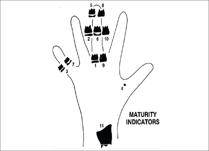
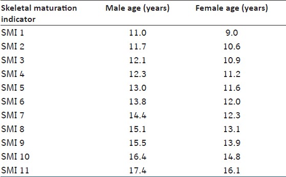
- Fishman [2] (1982): Hand-wrist radiographs for SMI as shown in [Figure 1]. Hand-wrist radiographs were assigned according to the standards given in the "Radiographic Atlas of Greulich and Pyle." [4]
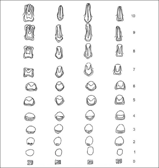
- C. M. Nolla [5] (1960): IOPA radiographs of maxillary and mandibular permanent right canine were assessed for dental age according to Nolla's [5] calcification stages [Figure 2].
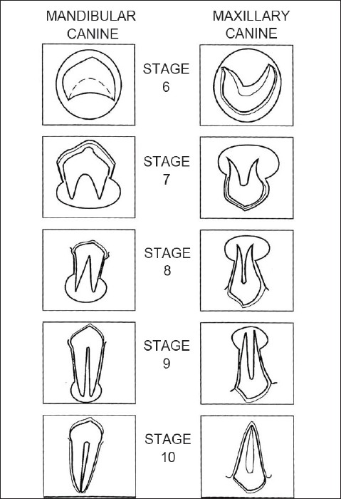 | Figure 2: Nolla's calcifi cation stages of maxillary and mandibular canine
Click here to view |
Nolla's developmental stages
The Nolla's developmental stages as shown in [Figure 3]. They are as follows:
stage 10: apical end of root completed
stage 9: root almost complete; open apex
stage 8: two-thirds of root completed
stage 7: one-third of root completed
stage 6: crown completed
stage 5: crown almost completed
stage 4: two-thirds of crown completed
stage 3: one-third of crown completed
stage 2: initial calcification
stage 1: presence of crypt
stage 0: absence of crown
Error of measurements
To evaluate the magnitude of error in the measurements of various stages of tooth development and skeletal maturation, repeated determination was carried out on 10 individuals at an interval of 15 days. These tracings were analyzed separately and two sets of reading were obtained from each case. The reliability of the measurements tested by "t" test was not found to be statistically significant.
Statistical analysis
Mean, standard deviation, and standard error were calculated for all the groups and correlation coefficients were computed for the samples collected. The Student "Newman-Keuls test" was employed to evaluate the difference between the mean values of chronological age and skeletal age, as assessed by skeletal maturation and canine calcification stages.
| Results | | |
[Table 2] shows age assessment as per Fishman's method.
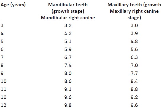
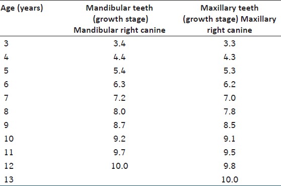
[Table 3] and [Table 4] show the norms for maturation of canines for boys and girls, respectively.
[Table 5] shows comparison of chronologic age with skeletal age assessed by SMI and calcification stages of maxillary and mandibular right canine. | Table 5: Comparison of chronologic age with age by SMI, age by Mandibular canine and age by maxillary canine
Click here to view |
[Table 6] shows correlation of age by SMI and by calcification stages of maxillary and mandibular right canine. | Table 6: Correlati on of age by SMI, age by max. Rt canine, and age by mand. Rt canine
Click here to view |
| Discussion | | |
In the present study, there was a significant difference between chronological age and skeletal age assessed by SMI. This supported the fact that skeletal maturation showed variation in comparison to chronological age. Hence, use of chronologic age to assess maturation status is questionable. This study was supported by a study done by Hunter, [6] which showed a significant difference between the mean chronological age and skeletal age. This study was also supported by Fishman [2] and Schour and Masseler. [7] There is insignificant difference between chronological age and age assessed by maxillary and mandibular right canine. This indicated that dental maturation in terms of development of canine also increased as chronological age. This study was supported by Anderson and coworkers [8] and Lemons and Gray. [9]
Correlation between age assessed SMI and canine calcification stages of maxillary and mandibular right canine was highly significant for males and females. This study was supported by Luterstein, [10] Lewis and Garn, [11] Sierra, [12] Coutinho and Bushchang, [13] Demirjian and Bushchang, [14] Green, [15] and Chertkow et al.[16] Considering this study, it could be stated that canine could also be used as an SMI.
As per this study, canine calcification stage 9 was related to capping of the third middle phalanx and appearance of the adductor sesamoid of the thumb. Hence, maxillary and mandibular canine calcification stage 9 confirmed the attainment of peak height velocity (PHV).
Intermediate stage of canine calcification between 8 and 9 could be used to identify the early stage of pubertal growth spurt. As these stages could be assessed on IOPA, this could prove more economical and convenient as armamentarium required is much simpler and even radiation dose is less.
It can be inferred that chronological age could not be used reliably for assessing skeletal maturity, but strong correlation was observed between SMI and maxillary and mandibular canine calcification stages. This confirmed the reliability and validity of canine calcification stages to be used as SMI. This also eliminated the use of additional radiographic exposure (hand-wrist radiograph) of patients in orthodontic practice because canine is recorded on panoramic radiograph.
In this study, females were ahead in skeletal maturation than males in all the age groups. [1] This is supported by Hagg and Taranger, [17] Castellanous et al, [18] Koshy and Tandon, [19] Prabhakar et al, [20] Hunter, [6] and Fishman. [2] Results of the present study show insignificant difference in dental development in males and females. The study was supported by Nolla. [5] The technique has the advantages of being simple, using low patient radiation dose, and exhibiting high degree of clarity of the radiographs. The equipment required is available in most dental clinics. For both the sexes, skeletal age (from hand-wrist) and dental age do not show high correlation with chronological age in all the age groups in this study. This indicates that the chronological age has no sufficient correlation with individual maturational development. Similar findings have been reported by Singer, [21] Demirjian et al[14] Prabhakar et al,[19] and Moorrees et al. [22]
To conclude, it could be stated that assessment of maturation is of utmost importance in certain orthodontic protocols like for myofunctional therapy, before starting with rapid maxillary expansion, and for timing of ortho-surgical procedures (surgery for mandibular setback should carried out only after mandibular growth has completed). As chronological age cannot show accurate status of individual's skeletal age, skeletal age could be assessed by time-tested hand-wrist radiographs or by canine calcification stages on periapical radiographs which are easier and cheaper to procure than hand-wrist X-rays. To further validate the results of this study, it should be carried out on larger sample size and varied age groups.
| Conclusion | | |
- Skeletal maturation was more advanced in comparison to chronological age in both males and females.
- There was good correlation between age assessed by SMI and canine calcification stages.
- Canine calcification stages could also be used as a skeletal maturity indicator besides SMI.
| References | | |
| 1. | Bala M, Pathak A, Jain RL. Assessment of skeletal age using MP3 and hand wrist radiograph and its correlation with dental age and chronological ages in children. J Indian Soc Pedod Prev Dent 2010;28:95-9. 
[PUBMED]  |
| 2. | Fishman LS. Maturational patterns and prediction during adolescence. Angle Orthod 1987;57:178-93.
|
| 3. | Fishman LS. Radoigarphic evaluation of skeletal maturation: A clinically oriented method based on hand wrist films. Angle Orthod 1982;52:88-112.
|
| 4. | Greulich WW, Pyle SI. Radiographic atlas of skeletal development of the hand and wrist. In: Todd TW, editor. Brush Foundation Study of Human Growth and Development initiated. California: Stanford University, Standford University Press; 1950.
|
| 5. | Nolla CM. The development of permanent teeth. J Dent Child 1960;27:254-66.
|
| 6. | Hunter CJ. The correlation of facial growth with body height and skeletal maturation at adolescence. Angle Orthod 1966;36:44-54.
|
| 7. | Schour, Masseler. The development of human dentition. J Am Dent Assoc 1941;281153-60.
|
| 8. | Anderson DL, Thompson GW, Popovitch F. Interrelationships of dental maturity, skeletal maturity, height and weight from age 4 to 14 years, Growth 1975;39:453-62.
|
| 9. | Lamons, Gray. A study of the relationship between tooth eruption age, skeletal development age and chronological age in sixty-one Atlanta children. Am J Orthod 1958;44: 687-91.
|
| 10. | Luterstein. A cross sectional study in dental development and skeletal age. J Am Dent Assoc 1961;62:161-7.
|
| 11. | Lewis, Garn. The relationship between tooth formation and other maturational factors. Angle Orthod 1960;30:69-77.
|
| 12. | Sierra AM. Assessment of dental and skeletal maturity: A new approach. Angle Orthod 1987;57:194-208.
|
| 13. | Coutinho, Buschang. Relationships between mandibular canine calcification stages and skeletal maturity. Am J Orthod 1993;104:262-8.
|
| 14. | Demirjian A, Buschang PH, Tanguay R, Patterson DK. Interrelationship among measures of somatic, skeletal, dental and sexual maturity. Am J Orthod 1985;88:433-8.
|
| 15. | Green LJ. Interrelationship among height, weight and chronological, dental and skeletal ages. Angle Orthod 1961;31:189-93.
|
| 16. | Chertkow S, Fatti P. The relationship between tooth mineralization and early radiographic evidence of adductor sesamoid calcification. Angle Orthod Angle Orthod 1979;49:282-8.
|
| 17. | Hagg U, Taranger J. Maturation indicators and the pubertal growth spurt. Am J Orthod 1982;82:299-309.
|
| 18. | Jiménez-Castellanos J, Carmona A, Catalina-Herrera CJ, Viñuales M. Skeletal maturation of wrist and hand ossification centers in normal spanish boys and girls: A study using the Greulich Pyle method. Acta Anat (Basel) 1996;155:206-11.
|
| 19. | Koshy S, Tandon S. Dental age assessment: The applicability of Demirjian's method in South Indian children. Forensic Sci Int 1998;94:73-85.
|
| 20. | Prabhakar AR, Panda AK, Raju OS. Applicablity of Demirjian's method of age assessment in children of Davangere. J Indian Soc Pedod Prev Dent 2002;20:54-62.
[PUBMED] |
| 21. | Singer J. Physiologic timing of orthodontic treatment. Angle Orthod 1980;50:322-33.
|
| 22. | Moorrees CF, Fanning EA, Hunt EE Jr. Age variation of formation stages for ten permanent teeth. J Dent Res 1963;42:1490-50.
|
[Figure 1], [Figure 2], [Figure 3]
[Table 1], [Table 2], [Table 3], [Table 4], [Table 5], [Table 6]
|


 Search Pubmed for