|
 
 |
|
REVIEW ARTICLE |
|
|
|
| Year : 2012 | Volume
: 18
| Issue : 3 | Page : 276-284 |
| |
Redefining the potential applications of dental stem cells: An asset for future
Shalu Rai1, Mandeep Kaur2, Sandeep Kaur1, Sapna Panjwani Arora1
1 Department of Oral Medicine and Radiology, Institute of Dental Studies and Technologies, Kadrabad, Modinagar, Uttar Pradesh, India
2 Department of Oral Medicine and Radiology, Dental College, Jamia Milia Islamia, New Delhi, India
| Date of Web Publication | 4-Mar-2013 |
Correspondence Address:
Shalu Rai
Department of Oral Medicine and Radiology, Institute of Dental Studies and Technologies, Kadrabad, Modinagar - 201 201, Uttar Pradesh
India
 Source of Support: None, Conflict of Interest: None  | 4 |
DOI: 10.4103/0971-6866.107976

 Abstract Abstract | | |
Recent exciting discoveries isolated dental stem cells from the pulp of the primary and permanent teeth, from the periodontal ligament, and from associated healthy tissues. Dental pulp stem cells (DPSCs) represent a kind of adult cell colony which has the potent capacity of self-renewing and multilineage differentiation. Stem cell-based tooth engineering is deemed as a promising approach to the making of a biological tooth (bio-tooth) or engineering of functional tooth structures. Dental professionals have the opportunity to make their patients aware of these new sources of stem cells that can be stored for future use as new therapies are developed for a range of diseases and injuries. The aim of this article is to review and understand how dental stem cells are being used for regeneration of oral and conversely nonoral tissues. A brief review on banking is also done for storing of these valuable stem cells for future use.
Keywords: Banking, dental stem cells, regeneration, stem cells from human exfoliated deciduous
How to cite this article:
Rai S, Kaur M, Kaur S, Arora SP. Redefining the potential applications of dental stem cells: An asset for future. Indian J Hum Genet 2012;18:276-84 |
How to cite this URL:
Rai S, Kaur M, Kaur S, Arora SP. Redefining the potential applications of dental stem cells: An asset for future. Indian J Hum Genet [serial online] 2012 [cited 2016 Jun 1];18:276-84. Available from: http://www.ijhg.com/text.asp?2012/18/3/276/107976 |
| Introduction | |  |
Stem cell research presents an opportunity for scientific evidence that goes far beyond regenerative medicine. Dentists are at the forefront of engaging their patients in potentially life-saving therapies derived from their own stem cells located either in deciduous or permanent teeth.
Two studies on research of human teeth in 2000 by The National Institute of Health (NIH) mentioned the discovery of adult stem cells in impacted third molars and even more resilient stem cells in deciduous teeth. [1] All the dental, oral, and craniofacial structures are formed by neural crest derived and/or mesenchymal cells during native development. Dental stem cells can be used in the regeneration of dentin and/or dental pulp; biologically viable scaffolds will be used for the replacement of orofacial bone and cartilage, and defective salivary glands will be partially or completely regenerated. [1]
Tooth banking is based on the firm belief that personalized medicine is the most promising avenue for treating challenging diseases and injuries that would occur throughout life. Individuals have different opportunities at different stages of their life for banking their valuable cells. Recent studies have shown that SHED have the ability to develop into more types of body tissues than other types of stem cells.
Two different populations of stem cells have been considered for tooth formation:
- Epithelial stem cells (EpSC), which will give rise to ameloblasts.
- Mesenchymal stem cells (MSC) that will form the odontoblasts, cementoblasts, osteoblasts, and fibroblasts of the periodontal ligament. [2]
| Epithelium Stem Cells | | |
Although significant progress has been made with MSC, there is no information available for dental epithelial stem cell (EpSC) in humans. There is an absence of dental epithelial stem cell (EpSC) populations in erupted teeth, precluding their use in tooth tissue engineering applications because their precursors are eliminated soon after eruption. [3] Stem cell technology appears to be the only possibility to recreate an enamel surface. [2]
| Mesenchymal Stem Cells | | |
The potential of dental MSC for tooth regeneration and repair has been extensively studied in the last years which can differentiate into chondrocytes, osteoblasts and adipocytes, and myocytes. [4] Hence, MSCs are more promising for therapeutic applications than other types of stem cells. [5]
Following types of mesenchymal progenitors have been assessed from teeth and have been used for regeneration of dental tissues. [6],[7]
Stem cells have been called DPSC (dental pulp stem cells), when found in permanent teeth, and SHEDs (stem cells from human exfoliated deciduous), when found in deciduous teeth in the periodontal ligament (periodontal ligament stem cells - PDLSCs). [6],[8]
Stem cells from human exfoliated deciduous teeth
These cells exhibited a high plasticity since they could differentiate into neurons, adipocytes, osteoblasts, and odontoblasts. In vivo SHED cells can induce bone or dentin formation. [3] SHEDs have a higher proliferation rate and these cells might represent a more immature population of multipotent stem cells. [9]
Adult dental pulp stem cells
Dental pulp stem cells (DPSCs) can be isolated from the dental pulp. [10] It has been shown that adult dental pulp contains precursors capable of forming odontoblasts under appropriate signals like calcium hydroxide or calcium phosphate materials. Tooth repair is a lifetime process, thus suggesting that MSC might exist in adult dental pulp. The in vivo therapeutic targeting of these adult stem cells remains to be explored. [2]
Periodontal ligament stem cells
PDL contains STRO-1 positive cells that maintain certain plasticity since they can adopt adipogenic, osteogenic, and chondrogenic phenotypes in vitro. According to Seo, et al.[11] it is thus obvious that PDL itself contains progenitors, which can be activated to self-renew and regenerate other tissues such as cementum and alveolar bone. [2],[10]
Stem cells from the apical part of the papilla
SCAP exhibit a higher proliferative rate and appears more effective than PDLSC for tooth formation. Importantly, SCAP are easily accessible since they can be isolated from human third molars. [2],[10]
Stem cells from the dental follicle
DFSC have been isolated from follicle of human third molars and express the stem cell markers Notch1, STRO-1, and nestin. Immortalized dental follicle cells are able to recreate a new periodontal ligament (PDL) after in vivo implantation. [2],[10]
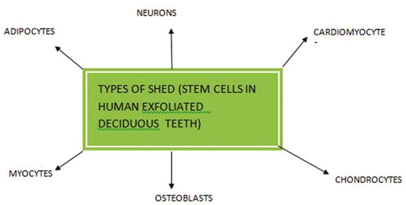
| Types of Stem Cells from Human Exfoliated Deciduous | | |
[Figure 1]
Adipocytes
Adipocytes have successfully been used to repair damage to the heart muscle caused by severe heart attack. There are also preliminary data to indicate that they can be used to treat cardiovascular disease, spine and orthopedic conditions, congestive heart failure, Crohn's disease, soft tissue grafts for facial soft tissue reconstruction and augmentation, [4] have the ability to repair damaged cardiac tissues following a heart attack, and can be used in plastic surgery. [12],[13],[14]
Chondrocytes and osteoblasts
Chondrocytes and osteoblasts have successfully been used to grow bone and cartilage suitable for transplant. They have also been used to grow intact teeth in animals. [12],[15],[16]
Mesenchymal
MSC-derived myocytes can be used to treat muscular dystrophy and facial muscle atrophy. Since they can form neuronal clusters, mesenchymal stem cells have the potential to treat neuronal degenerative disorders such as Alzheimer's and Parkinson's diseases, cerebral palsy, as well as a host of other disorders. [12],[15],[16],[17],[18]
SHED and banking
Existing research has clearly shown that primary teeth are a better source for therapeutic stem cells than wisdom teeth, and orthodontically extracted teeth. [16] With the documented discovery of SHED in 2003 by Dr. Shi, [15] an accessible and available source of stem cells has been identified which can be easily preserved and used for future cure of ailments. SHED are immature, unspecialized cells in the teeth that are able to grow into specialized cell types by a process known as "differentiation." Abbas, et al.[19] investigated and stated that SHED are of neural crest origin. [12]
The main advantage of banking SHED cells is that it provides a guaranteed matching donor (autologous transplant) for life and saves cells before natural damage occurs. Apart from these, the other advantages are as follows:
- Simple and painless for both child and parent and less than one third of the cost of cord blood storage. [12]
- SHED cells are complementary to stem cells from cord blood. [16]
- SHED may also be useful for close relatives of the donor. [5]
Also these are adult stem cells and thus are not the subject of same ethical concerns as embryonic stem cells. [5],[20]
Collection, isolation and preservation of SHED
For deciduous teeth, the best candidates for isolation are canine and incisors with the presence of healthy pulp that are starting to loosen. In children, other sources for easily accessible stem cells are supranumerary teeth, mesodens, overretained deciduous teeth associated with congenitally missing permanent teeth and prophylactic removal of deciduous molars for orthodontic indications. When a deciduous tooth becomes extremely mobile it is likely that the pulp has been separated from its blood supply. The tooth might still maintain its gingival attachment and be retained for weeks in the mouth with a necrotic pulp. Adolescents have two excellent opportunities for banking their stem cells from extracted teeth: following extraction of bicuspid teeth for orthodontic treatment and when their wisdom teeth are extracted. The follicular sac of an unerupted tooth may also prove to be a valuable source for stem cells. [5]
As such, it is inevitable that the key to successful stem cell therapy lies in being able to harvest the cells at the right point of development and to safely store them until accident or disease requires their usage. It is needless to say that this means potentially storing for decades, and the cost and technical difficulty of doing this properly make stem cell therapy using one's own cells a still uncertain bet. Tooth banking is not very popular but the trend is catching up mainly in the developed countries. [12]
Step 1: Tooth collection
With prior informed consent, the first step is to place the tooth in a sterile saline solution [12] or in fresh milk in the storage container along with frozen gel packs. The kit is then ready for delivery to their lab. [21] The tooth exfoliated should have pulp which is red in color, and not necrotic, thus indicating that the pulp received blood flow till the time of removal, which is indicative of cell viability. After its recovery, the tooth is transferred into the vial containing a hypotonic phosphate buffered saline solution, which provides nutrients and helps to prevent the tissue from drying out during transport (up to four teeth in the one vial). The vial is then carefully sealed and placed into the thermette, a temperature phase change carrier, which is then placed into an insulated metal transport vessel. This procedure maintains the sample in the hypothermic state during transportation and is described as sustentation. [12]
Step 2: Stem cell isolation [12]
When the tooth bank receives the kit or vial, all the cells are isolated and the stringent protocol is followed for cleaning of the tooth surface by various disinfectants; isolation of pulp tissue from pulp chamber and cells is then cultured in a mesenchymal stem cell medium (MSC) under appropriate conditions. By making changes in the MSC medium different cell lines can be obtained such as odontogenic, adipogenic, and neural. If cultures are obtained with unselected preparation, colonies of cells with morphology resembling epithelial cells or endothelial cells can be established.
Usually cells disappear during the course of successive cell passages. If contamination is extensive, then a change in procedures can be performed: in which STRO-1 or CD 146 can be used. This is considered as most reliable.
The time from harvesting to arrival at the processing storage facility should not exceed 40 hours.
Step 3: Stem cell storage
In the light of present research, either of the following two approaches is used for stem cell storage:
- Cryopreservation
- Magnetic freezing
Cryopreservation [4],[11],[21],[22]
Cells are preserved by cooling them to subzero temperatures, at which biological activity is stopped. The cells are preserved in a liquid nitrogen vapor at a temperature of less than −150° C. This preserves the cells and maintains their latency and potency.
Magnetic freezing [12],[23]
Hiroshima University uses magnetic freezing rather than cryogenic freezing. The idea of this technique is to completely chill an object below the freezing point, by using a magnetic field, without freezing, thus ensuring, distributed low temperature without the cell wall damage caused by ice expansion and nutrient drainage due to capillary action, as normally caused by conventional freezing methods. Then, once the object is uniformly chilled, the magnetic field is turned off and the object snap freezes.
Using CAS, Hiroshima University claims that it can increase the cell survival rate in teeth to a high of 83%. This system is a lot cheaper than cryogenics and more reliable as well. [2]
Licensed tooth banks are the following: [24]
- Reliance life sciences, Delhi.
- Life call, Chennai.
- Stemade Biotech Pvt Ltd. India.
- The Norwegian Tooth Bank.
Dental tissue regeneration
The goal of tissue engineering is to restore tissue function through the delivery of stem cells, bioactive molecules, natural/synthetic tissue support or scaffold to build a three-dimensional living construct that is functionally, structurally and mechanically equal to or better than the tissue that is to be replaced. [25]
Currently there are two major approaches to tooth regeneration:
- By using principles of tissue engineering.
- By reproducing the developing processes of embryonic tooth formation. [26]
By using principles of tissue engineering Yelick's group have used dental epithelium and pulpal mesenchymal tissues and seeded the mixture of heterogenous single cells onto tooth-shaped biodegradable polymer scaffold consisting of polyglycolic acid and polycoglycolide copolymer resulting in tiny tooth like tissues (such as enamel, dentin, pulp) within implant, which resembled the crown of natural teeth after 25-30 weeks of placement of this cell scaffold construct into the body of animal host. Further studies are required to achieve structurally sound teeth as the success rate of natural tooth like looking teeth is 15-20%. [27]
Developing processes of embryonic tooth formation
Hu, et al.[28] have shown that under experimental conditions, nondental mesenchymal cells, i.e., bone marrow cells rather than dental mesenchymal cells, can give rise to ameloblast-like cells that form enamel while Sharpe's group [29] reported that bone marrow cells when placed in contact with oral epithelium possess the potential to differentiate into dental mesenchymal cells forming dentin and pulp.
Root regeneration
Current understanding of the cellular and molecular mechanisms of tooth root development is still in its infancy, although a number of genes involved in tooth crown development have been identified. Sonoyama, et al.[30] explored the potential for reconstructing a functional tooth in miniature pigs in which a bio-root periodontal complex is built up by postnatal stem cells using stem cells from apical root papilla (SCAP) [31] and periodontal ligament stem cells to which an artificial porcelain crown is fixed and concluded that dental stem cell engineering can be used for human tooth root regeneration. [30]
Limitations in tooth regeneration research
Certain limitations are listed below as suggested by Nakahara and Idle: [32]
- Will the principles of tissue engineering related to tooth regeneration be able to resemble correct tooth morphology or not?
- There is no suitable substitute for the embryonic oral epithelium even though adult bone marrow cells can replace dental mesenchymal cells and at the present time there is no embryonic environment that enables bone marrow cells to differentiate into tooth germ cells.
- Concern over host immune rejection and ethical issue on use of human embryo still exists till date.
Dental stem cells for regeneration of nondental tissues [Figure 2]
Fang, et al.[33] in 2007 demonstrated that bone marrow MSCs were capable of generating bone structures and bone-associated marrow elements on the surfaces of the orofacial bone. In addition, they found that the subcutaneous transplantation of another population of MSCs, the human periodontal ligament stem cells (PDLSCs), could form substantial amounts of collagen fibers and improve facial wrinkles in mouse. [34]
Otaki, et al.[35] in 2007 reported that dental pulp cells (first to eighth passage) produced bone instead of dentin when those cells were implanted into subcutaneous sites in immunocompromised mice with HA/TCP powder as their carrier. Their evidence showed that dental pulp cells are the common progenitors of odontoblasts and osteoblasts, or dental pulp cells are mesenchymal stem cells themselves. | Figure 2: Uses of dental stem cells in regeneration of nondental tissues
Click here to view |
Nosrat, et al.[36] in 2001 established an in vitro culture system to study the interactions between the dental pulp cells and trigeminal neurons. When DPC are cocultured with trigeminal neurons, they promote survival and a specific and elaborate neurite outgrowth pattern from trigeminal neurons, whereas skin fibroblasts do not provide a similar support. Interestingly, grafting the dental pulp tissue into hemisected spinal cord increases the number of surviving motoneurons, indicating a functional bioactivity of the dental pulp-derived neurotrophic factors in vivo by rescuing motoneurons.
Application of dental stem cells in dentistry.
Application of dental stem cell in various fields of dentistry has been explored and has been mentioned briefly [Figure 3].
Regenerative endodontic techniques
These techniques are based on the basic tissue engineering principles. [37],[38],[39],[40]
Murray, et al.[37] identified several major areas of research that might have applications in the development of these techniques.
These techniques are as follows:
- Dentin regeneration by pulp implantation.
- Revascularization of root canal.
- Three-dimensional cell printing.
- Gene delivery-based approaches.
- Postnatal stem cell therapy.
- Scaffold implantation and delivery.
Cultured cells from tooth bud tissue were then harvested and seeded onto biodegradable polyester scaffolds, the purpose of which was to provide a support onto which seeded dental progenitor cells - postnatal dental stem cells (PNDSC) [41] - can adhere and orient themselves with respect to each other, allowing for requisite epithelial and mesenchymal dental cell interactions for tooth initiation and development. [2]
Damaged odontoblasts can be replaced by newly generated populations of odontoblasts, [42] derived from stem cells from pulp and regenerate dentine/pulp tissue [43],[44] resembling normal physiologic tissue. [45],[46],[47]
Several case reports have documented revascularization [48],[49] of necrotic root canal systems of immature permanent teeth by disinfection followed by establishing bleeding into the canal system [31] via little or no instrumentation to avoid fracture of thin dentinal wall.
The formation of a blood clot might serve as a protein scaffold, permitting three-dimensional ingrowth of tissue [37],[50],[51] and both dentin and platelets provide sources of certain growth factors. [36]
The three-dimensional cell printing technique can be used to precisely position cells, and this method has the potential to create tissue constructs that mimic the natural tooth pulp tissue structure. However, early research has yet to show that three-dimensional cell printing can create functional tissue in vivo. [37]
The major reason for using stem cells in cell-based gene therapies is that they are a self-renewing population of cells and thus may reduce or eliminate the need for repeated administrations of the gene therapy. The clinical applications of hematopoietic stem cell-based gene therapies are diverse, extending to organ transplantation, blood and bone marrow disorders, and immune system disorders. [45],[52] Both in vivo and ex vivo approaches can be used for gene therapy. The ex vivo approach involves genetic manipulation of cells in vitro, which are subsequently transplanted to the regeneration site. The successful bone induction has been reported after application of the BMP family members, Bmp2, Bmp4, Bmp7, and Bmp9 by gene therapy using viral vectors. Rutherford [53] in his work used ferret pulps with cDNA-transfected mouse BMP-7 but failed to produce reparative response, suggesting that further research in the potential of pulp gene therapy is needed.
Periodontal regeneration
Recent studies revealed the presence of progenitor cells in the dental follicle, which can contribute to the formation of periodontal tissues (including cementum and the periodontal ligament) known as Dental follicle stem cells and odontoblasts. Kramer et al.[54] demonstrated that PDL-like tissue can be developed from periodontal progenitor cells and from mesenchymal stem cells in contact with either PDL factors or the tissue itself. Saito et al.[55] suggested the use of bone morphogenetic protein BMP-2 as a promoter for the differentiation of progenitor cells from the dental follicle into cementoblasts and odontoblasts in order to reestablish the integrity of the PDL. [45]
The potential use of bone-marrow-derived MSCs (BMSSCs) for the regeneration of dental structures was tested in a study by Kawaguchi and colleagues [56] where autologous cells were transplanted into surgically created dental defects in beagle dogs in experimental class III furcation defects. Newly formed cementum was detected covering the surface of the roots and the presence of Sharpey's fibers inserted into the cementum was noted. [57]
Bone and craniofacial regeneration
Stem cells have been used in the tissue engineering of a human-shaped temporomandibular joint. MSCs were first isolated from bone marrow and exposed separately to either chondrogenic or osteogenic supplemented culture medium. [1],[4] Alhadlaq and Mao [58],[59] used MSC-derived cells encapsulated in a poly {ethylene glycol} diacrylate (PEGDA) hydrogel that was molded into an adult human mandibular condyle in stratified yet integrated layers of cartilage and bone. The osteochondral grafts in the shape of human TMJs were implanted in immunodeficient mice for up to 12 weeks. Upon harvest, the tissue-engineered mandibular joint condyles retained their shape and dimensions. [1]
According to Pittenger, et al.[60] bone marrow-derived MSC are now under consideration for the repair of craniofacial bone and even the replacement or regeneration of oral tissues. [1],[61] Reconstruction of craniofacial and dental defects using MSC avoids many of the limitations of both auto- and allografting techniques. [62] Clinical studies are being conducted using stem cells for alveolar ridge augmentation and long-bone defects. [63],[64] Vascularized bone grafts are also in development using stem cells, and reconstruction of a patient's resected mandible has been carried out using this technique. [1]
In study by Alejandro, et al.[65] the authors used osteogenically differentiated fat-derived stem cells to repair surgically created rat palatal bone defects. Hematoxylin and eosin staining, immunohistochemical staining for osteocalcin, and histomorphometric measurements of new bone were performed. Newly formed bone stained positive for osteocalcin.
Changes in the expression of stem cell markers in oral lichen planus and hyperkeratotic lesions
Despite the pivotel role of stem cells in homeostasis of oral epithelial the location of this cell population within the tissue is uncertain. How disease influences these cells in vivo also remains to be elucidated.
Kose, et al.,[66] in a study showed the location of stem cells in normal nonkeratinised buccal epithelium (NOM) by immunohistochemical staining for the putative stem cell markers alpha 6 and beta 1 integrins, melanoma-associated chondroitin sulphate proteoglycan (MCSP), NG2 the rat homologue of human MCSP, notch 1 and keratin 15 (k15). This is the first study to show alterations in stem cell marker expression in oral lichen planus (OLP) and oral hyperkeratotic lesions (OHK) which indicates pathological signaling may regulate expression of these markers. This implicates adult stem cells in the pathogenesis of these mucosal disorders where epithelial differentiation and proliferation is known to be perturbed. k15, NG2 and beta 1 staining was continuous in the basal layer of NOM while α6 and β1 and MCSP were upregulated in both OLP and OHK. NG2 remained unchanged and notch 1 was absent in all samples. Therefore, the stem cell phenotype in OLP and OHK may be altered in response to pathological signaling. Classification of these changes is essential to understand the role of adult stem cells in the pathogenesis of oral diseases characterized by abnormal keratinocyte proliferation and differentiation.
Staining pattern observed in the study showed that α6 integrin and MCSP are the most likely adult stem cell markers in oral epithelium. High expression of the integrin family of receptors is believed to be responsible for stem cells being more adhesive to basement membrane than other basal cells. Further studies of these molecular perturbations are essential to understand the fundamental role of adult stem cells in the pathogenesis of benign mucosal disease.
| Conclusion | | |
The current research on dental stem cells is expanding at an unprecedented rate. At present, stem-cell-based tissue engineering approaches provide the most promising solution. Within the next few years, these cells will be used to restore the form and function of the oral cavity using autologous cells, thereby overcoming histocompatability mismatch and transmission of viral disease. While we can see the promise of human stem cell therapies for the future, dentists know that it is important to act now to harvest and store these mesenchymal stem cells from deciduous teeth and extracted permanent teeth because the opportunity to bank patient's dental stem cells will have greatest future impact if seized while patients are young and healthy for future regenerative therapies, although there are many ethical controversies and legal and social questions which need to be addressed before stem cell-based therapies become clinically available.
| References | | |
| 1. | Mao JJ, Collins FM. Stem Cells: Sources, Therapies and the Dental Professional. Available from: http://www.ineedce.com/courses/1486/PDF/StemCells.pdf. [Last accessed on 2011 May 10]. 
|
| 2. | Bluteau G, Luder HU, Bari DE, Mitsiadis TA. Stem cells for tooth engineering. Eur Cell Mater 2008;16:1-9.
|
| 3. | Yelick PC, Vacanti JP. Bioengineered teeth from tooth bud cells. Dent Clin N Am 2006;50:191-203.
|
| 4. | Mao JJ. Stem cell and Future of Dental Care. N Y State Dent J 2008;74:20-4.
|
| 5. | Reznick JB. Stem Cells: Emerging medical and dental therapies for the dental professional. October 2008. Available from: http://www.stemsave.com/Docs/News/Dentaltown.com. [Last accessed on 2011 May 02].
|
| 6. | Nedel F, André Dde A, de Oliveira IO, Cordeiro MM, Casagrande L, Tarquinio SB, et al. Stem Cells: Therapeutic Potential in Dentistry. J Contemp Dent Pract 2009;10:90-6.
|
| 7. | Peneva M, Mitev V, Ishketiev N. Isolation of mesenchymal stem cells from the pulp of deciduous teeth. Journal of IMAB -
|
| 8. | Annual Proceeding (Scientific Papers) 2008;2:84-7.
|
| 9. | D′Aquino R, De Rosa A, Laino G, Caruso F, Guida L, Rullo R, et al. Human dental pulp stem cells: From biology to clinical applications. J Exp Zool B Mol Dev Evol 2009;312B: 408-15.
|
| 10. | Abukawa H, Papadaki M, Abulikemu M, Leaf J, Vacanti JP, Kaban LB, et al. The engineering of craniofacial tissues in the laboratory: A review of biomaterials for scaffolds and implant coatings. Dent Clin N Am 2006;50:205-16.
|
| 11. | Ulmer FL, Winkel A, Kohorst P, Stiesch M. Stem cells -Prospects in dentistry. Schweiz Monatsschr Zahnmed 2010;120:860-83.
|
| 12. | Seo BM, Miura M, Sonoyama W, Coppe C, Stanyon R, Shi S. Recovery of stem cells from cryopreserved periodontal ligament. J Dent Res 2005;84:907-12.
|
| 13. | Arora V, Arora P, Munshi AK. Banking stem cells from human exfoliated deciduous teeth (SHED): Saving for the Future. J Clin Pediatr Dent 2009;33:289-94.
|
| 14. | Gandia C, Armiñan A, García-Verdugo JM, Lledó E, Ruiz A, Miñana MD, et al. Human dental pulp stem cells improve left ventricular function, induce angiogenesis, and reduce infarct size in rats with acute myocardial infarction. Stem Cells 2007;26:638-45.
|
| 15. | Perry BC, Zhou D, Wu X, Yang FC, Byers MA, Chu TM, et al. Collection, cryopreservation, and characterization of human dental pulp-derived mesenchymal stem cells for banking and clinical use. Tissue Eng Part C Methods 2008;14:149-56.
|
| 16. | Miura M, Gronthos S, Zhao M, Lu B, Fisher LW, Robey PG, et al. SHED: Stem cells from human exfoliated deciduous teeth. Proc Natl Acad Sci U S A 2003;100:5807-12.
|
| 17. | Shi S, Bartold PM, Miura M, Seo BM, Robey PG, Gronthos S. The efficacy of mesenchymal stem cells to regenerate and repair dental structures. Orthod Craniofac Res 2005;8:191-9.
|
| 18. | Arthur A, Rychkov G, Shi S, Koblar SA, Gronthos S. Adult human dental pulp stem cells differentiate toward functionally active neurons under appropriate environmental cues. Stem Cells 2008;26:1787-95.
|
| 19. | Kerkis I, Ambrosio CE, Kerkis A, Martins DS, Zucconi E, Simone Fonseca AS, et al. Early transplantation of human immature dental pulp stem cells from baby teeth to golden retriever muscular dystrophy (GRMD) dogs: Local or systemic? J Transl Med 2008;6:35.
|
| 20. | Abbas A, Diakonov I, Sharpe P. Neural Crest Origin of Dental Stem Cells. Pan European Federation of the International Association for Dental Research (PEF IADR). Seq #96-Oral Stem Cells: Abs, 0917, 2008.
|
| 21. | Mao JJ. Stem Cells and the Future of Dental Care. N Y State Dent J 2008;74:21-4.Available from: http://www.BangkokDentalHospital.com/stem cell/tooth stem cells. [Last accessed on 2011 May 10].
|
| 22. | Woods EJ, Perry PE, Hockema JJ, Larson L, Zhou D, Goebel WS. Optimized cryopreservation method for human dental pulp-derived stem cells and their tissues of origin for banking and clinical use. Cryobiology 2009;59:150-7.
|
| 23. | TT-450-Stem Cells and Teeth Banks, ebiz news from Japan. Available from: http://www.japaninc.com/tt450. [Last accessed on 2011 May 01].
|
| 24. | Umbilical cord Blood Banking [Internet]. Available from: http://www.chillibreeze.com. [Last accessed on 2011 May 03].
|
| 25. | Taba M Jr, Jin Q, Sugai JV, Giannobile WV. Current concept in periodontal bioengineering. Orthod Craniofacial Res 2005;8:292-302.
|
| 26. | Dadu SS. Tooth regeneration current status. Indian J Dent Res 2009;20:506-7.
[PUBMED]  |
| 27. | Young CS, Terada S, Vacanti JP, Honda M, Bartlett JD, Yelick PC. Tissue Engineering of Complex Tooth Structures on Biodegradable Polymer Scaffolds. J Dent Res 2002;81:695-700.
|
| 28. | Hu B, Unda F, Kuchler SB, Jimenez L, Wang XJ, Haïkel Y, et al. Bone marrow cells can give rise to ameloblast-like cells. J Dent Res 2006;85:416-21.
|
| 29. | Ohazama A, Modino SA, Miletich I, Sharpe PT. Stem-cell-based tissue engineering of murine teeth. J Dent Res 2004;83:518-22.
|
| 30. | Sonoyama W, Liu Y, Fang D, Yamaza T, Seo BM, Zhang C, et al. Mesenchymal stem cell-mediated functional tooth regeneration in swine. PLoS ONE 2006;1:e79.
|
| 31. | Huang GT . The hidden treasure in apical papilla: The potential role in pulp/dentin regeneration and bioroot engineering. J Endod 2008;34:645-51.
|
| 32. | Nakahara T, Ide Y. Tooth regeneration: Implications for the use of bioengineered organs in first-wave organ replacement. Human Cell 2007;20:63-70.
|
| 33. | Fang D, Seo BM, Liu Y, Sonoyama W, Yamaza T, Zhang C, et al. Transplantation of mesenchymal stem cells is an optimal approach for plastic surgery. Stem Cells 2007;25:1021-8.
|
| 34. | Science behind Dental Pulp Stem Cells. Available from: http://www.ncbi.nlm.nih: gov/entrez/query/pubmed and cmd/search and term 17170063 [Last accessed on 2011 May 05].
|
| 35. | Otaki S, Ueshima S, Shiraishi K, Sugiyama K, Hamada S, Yorimoto M, et al. Mesenchymal progenitor cells in adult human dental pulp and their ability to form bone when transplanted into immunocompro- mised mice. Cell Biol Int 2007;31:1191-7.
|
| 36. | Nosrat IV, Widenfalk J, Olson L, Nosrat CA. Dental pulp cells produce neurotrophic factors, interact with trigeminal neurons in vitro, and rescue motoneurons after spinal cord injury. Dev Biol 2001;238:120-32.
|
| 37. | Murray PE. Regenerative endodontics: A review of current status and a call for action. J Endod 2007;33:377-90.
|
| 38. | Hargreaves KM, Giesler T, Henry M, Wang V. Regeneration potential of the young permanent tooth: What does the future hold? J Endod 2008;34:S51-6.
|
| 39. | Cohen S. The Transformation of Endodontics in the 21 st Century. Smile Dent J 2010;5:1-7.
|
| 40. | Zhang W, Yelick PC. Vital pulp therapy-Current progress of dental pulp regeneration and revascularization. Int J Dent 2010;2010:1-9.
|
| 41. | Sloan AJ, Smith AJ. Stem cells and the dental pulp: potential roles in dentine regeneration and repair. Oral Dis 2007;13:151-7.
|
| 42. | Yu JD, Deng Z, Shi J, Zhai H, Nie X, Zhuang H, et al. Differentiation of dental pulp stem cells into regular-shaped dentin-pulp complex induced by tooth germ cell conditioned medium. Tissue Engg 2006;12:3097-105.
|
| 43. | Simon S, Cooper P, Berdal A, Lumley P, Tomson P, Anthony JS. Understanding pulp biology for routine clinical practice. ENDO (Lond Engl) 2009;3:171-84.
|
| 44. | Smith AJ, Lumley PJ, Tomson PL, Cooper PR. Dental regeneration and materials: A partnership. Clin Oral Invest 2008;12:103-8.
|
| 45. | Nakahara T. A review of new developments in tissue engineering therapy for periodontitis. Dent Clin N Am 2006;50:265-76.
|
| 46. | Nakashima M, Iohara K. Pulp stem cells and pulp regeneration. Tissue injury and pulp regeneration. IADR Pulp Biology and Regeneration Group Symposium Geneva, 2010. p. 25.
|
| 47. | El-Backly RM, Massoud AG, El-Badry AM, Sherif RA. Regeneration of dentine/pulp-like tissue using a dental pulp stem cell/poly (lactic-co-glycolic) acid scaffold construct in New Zealand white rabbits. Aust Endod J 2008;34:52-67.
|
| 48. | Markowicz M, Koellensperger M, Neuss S, Steffens GC, Pallua N. Enhancing the vascularization of three-dimensional scaffolds: new strategies in tissue regeneration and tissue engineering. Topics in Tissue Engineering 2005;2:1-15.
|
| 49. | Shin SY, Albert JS, Mortman RE. One step pulp revascularization treatment of an immature permanent tooth with chronic apical abscess: A case report. Int Endod J 2009;42:1118-26.
|
| 50. | Reynolds K, Johnson JD, Cohenca N. Pulp revascularization of necrotic bilateral bicuspids using a modified novel technique to eliminate potential coronal discolouration: A case report. Int Endod J 2009;42:84-92.
|
| 51. | Trope M. Regenerative potential of dental pulp. J Endod 2008;34 (7 Suppl):S13-7.
|
| 52. | Use of genetically modified stem cells in experimental gene therapies. Stem cell information. The National Institutes of Health Resource for stem cell research. Available from: http://stemcells.nih.gov/info/scireport/chapter 11. [Last accessed on 2011 May 12].
|
| 53. | Rutherford RB. BMP-7 gene transfer to inflamed ferret dental pulps. Eur J Oral Sci 2001;109:422-4.
|
| 54. | Kramer PR, Nares S, Kramer SF, Grogan D, Kaiser M. Mesenchymal Stem Cells Acquire Characteristics of Cells in the Periodontal Ligament in vitro. J Dent Res 2004;83:27-34.
|
| 55. | Saito N, Takaoka K. New synthetic biodegradable polymers as BMP carriers for bone tissue engineering. Biomaterials 2003;24:2287-93.
|
| 56. | Hiroyuki K, Aakio H, Naohiko H, Tomoyuki I, Hidenori H, Hideki S, et al. Enhancement of periodontal tissue regeneration by transplantation of bone marrow mesenchymal stem cells. J Periodontol 2004;75:1281-7.
|
| 57. | Goldberg M, Lacerda-Pinheiro S, Jegat N, Six N, Septier D, Priam F, et al. The impact of bioactive molecules to stimulate tooth repair and regeneration as part of restorative dentistry . Dent Clin North Am 2006;50:277-98.
|
| 58. | Alhadlaq A, Mao JJ. Mesenchymal stem cells: Isolation and therapeutics. Stem Cells Dev 2004;13:436-48.
|
| 59. | Alhadlsq A, Mao JJ. Tissue-engineered neogenesis of human-shaped mandibular condyie from rat mesenchymal stem cells. J Dent Res 2003;82:951-6.
|
| 60. | Pittenger MF, Mackay AM, Beck SC, Jaiswal RK, Douglas R, Mosca JD, et al. Multilineage potential of adult human mesenchymal stem cells. Science 1999;284:143-7.
|
| 61. | Wan DC, Nacamuli RP, Longaker MT. Craniofacial Bone Tissue Engineering. Dent Clin North Am 2006;50:175-90.
|
| 62. | Iohara K, Nakashima M, Ito M, Ishikawa M, Nakasima A, Akamine A. Dentin regeneration by dental pulp stem cell therapy with recombinant human bone morphogenetic protein 2. J Dent Res 2004;83:590-5.
|
| 63. | Ueda M, Yamada Y, Orawa R, Oluzaki Y. Clinical case reports of injectable tissue-engineered bone for alveolar augmentation with simultaneous implant placement. Int J Periodontics Restorative Dent 2005;25:129-37.
|
| 64. | Hibi H, Yamada Y, Ueda M, Endo Y. Alveolar cleft osteoplasty using tissue-engineered osteogenic material. Int I Oral Maxillofac Surg 2006;35:551-5.
|
| 65. | Alejandro CJ, James A. Repair of palatal bone defects using osteogenically differentiated fat-derived stem cells. Plast Reconstr Surg 2006;117:857-63.
|
| 66. | Kose O, Lalli A, Kutulola AO, Odell EW, Waseem A. Changes in the expression of stem cell markers in oral lichen planus and hyperkeratotic lesions. J Oral Sci 2007;49:133-9.
|
[Figure 1], [Figure 2], [Figure 3]
| This article has been cited by | | 1 |
Percepcija znanstvenika i poslijediplomskih studenata o budućnosti matičnih stanica u stomatologiji | [Perception of dental scientists and post-graduate students regarding future prospects of stem cells in dentistry] |
|
| Nagraj, A., Acharya, S. | | Acta Stomatologica Croatica. 2013; 47(4): 312-321 | | [Pubmed] | |
|
|
|
|