Targeted Delivery of Radioprotective Agents to Mitochondria
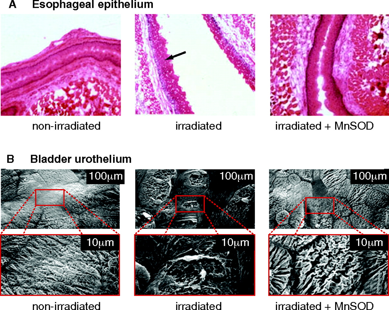
Radiation damage and protection by MnSOD. A. Brightfield micrograph depicting changes that occurred in esophageal epithelium after irradiation (30 Gy). The non-irradiated esophagus showed normal intact epithelium (n=5). Alternatively, the arrow in the irradiated micrograph points to holes in esophageal epithelial cells which represent nuclei that have undergone apoptosis and are no longer present (n=5). Intraesophageal administration of MnSOD 24 hours prior to irradiation protected the epithelial cells from undergoing apoptosis (n=5). Adapted from (35). B. Scanning electron micrographs depicting changes that occurred in the bladder urothelium after irradiation. The bottom electron micrographs are enlargements of the areas enclosed by the rectangles in the top micrographs. The non-irradiated bladders showed normal intact urothelium (n=7). However, 48 hours after irradiation (35 Gy), the bladder urothelium showed areas of superficial ulcerations of the umbrella cells (n=6). In rat bladders transfected intravesically with the human MnSOD transgene 24 hours prior to irradiation, the urothelium showed only minimal ulcerations (n=5). Adapted from (22, 37).

