- Institution: Stanford Univ Med Ctr Lane Med Lib/Periodical Dept/Rm L109
- Sign In as Member / Individual
MOSAICISM OF THE RETINAL PIGMENT EPITHELIUM: seeing the small picture
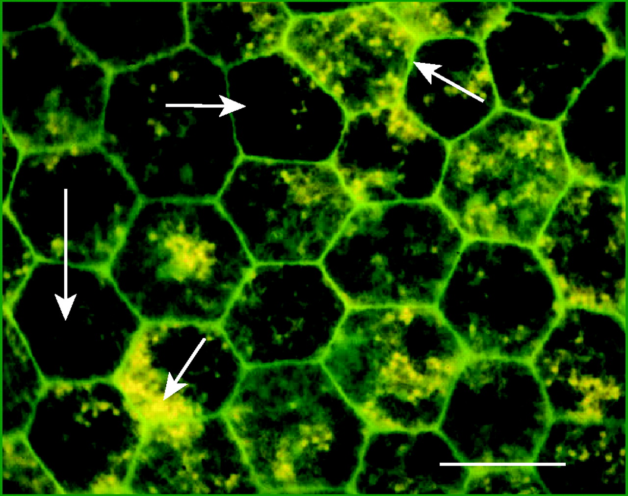
Figure 1.
Whole mount of the human RPE monolayer stained with fluorescein phalloidin for actin to show similarity in cell shape but heterogeneity in granule content. Actin is organized in a circumferential microfilament bundle (green), which illustrates the regular hexagonal shape of the cells. Individual cells vary in granule content, with some cells having abundant brown pigment granules (large arrows) and others abundant yellow lipofuscin granules (small arrows). The specimen was prepared by lifting off the neural retina, then processing the RPE with underlying choroid for epifluorescence microscopy. Scale bar: 20 μm.

