Finding Homes for Orphan Cytochrome P450s: CYP4V2 and CYP4F22 in Disease States
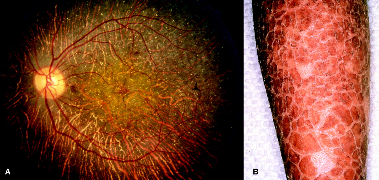
Figure 1
Disease states associated with mutations of genes that encode CYP4 enzymes
A. Fundus photograph of the eye of a Bietti’s patient illustrating typical yellow crystalline deposits and atrophy of the retinal pigmented epithelium. [From (13), with permission.] B. Hyperkeratotic scaling on the leg of a lamellar ichthyosis patient. (Courtesy of Dr. Philip Fleckman, Department of Dermatology, University of Washington, Seattle.)

