


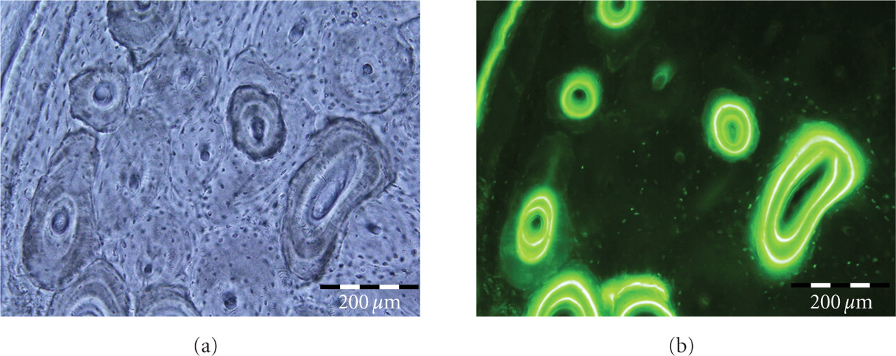
Figure 2
Pair of images (a) Brightfield and (b) Epifluorescent of cortical bone from a dog femur. Osteonal architecture of bone is evident. In addition labeled and unlabeled osteons can be clearly distinguished. The reversal lines and bone labels clearly demarcated the labeled and unlabeled osteons.