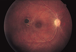
Figure 4. Three months after initial evaluation, there is retinovascular narrowing, central macular pigment change, mild optic nerve pallor, and a new chorioretinal scar in the region of the retinal infiltrate seen in Figure 3. Visual acuity was 20/25 OD.