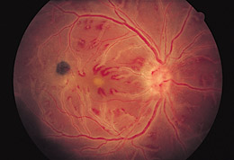
Figure 1. Fundus photograph of the right eye shows diffuse sheathing of all retinal vessels, perivenular hemorrhages, serous macular detachment, and a pigmented chorioretinal scar. Visual acuity was counting fingers with a 3+ relative afferent pupillary defect. Visual acuity was 20/20 OS.